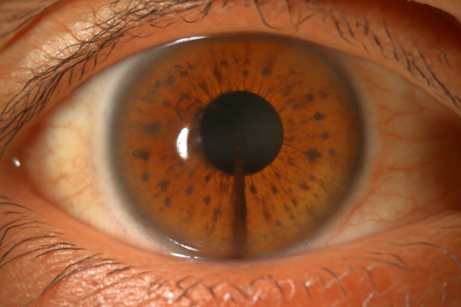
Overview and clinical significance of pigment dispersion syndrome
Definition and key features
Pigment dispersion syndrome travels quietly within the eye, with pigment shed from the posterior iris into the anterior chamber. Often asymptomatic, its clinical significance lies in the risk of raised intraocular pressure and pigmentary glaucoma. Routine exams can reveal this condition before symptoms appear!
Definition and key features include the following indicators:
- Posterior iris concavity with pigment release
- Pigment on the corneal endothelium (visible as a Krukenberg spindle)
- Mid-peripheral iris transillumination defects
- Potential intraocular pressure rise and glaucoma risk
- Common in younger, myopic eyes
These features inform monitoring strategies in South Africa’s eye clinics.
Symptoms and signs to watch for
pigment dispersion syndrome hides in the quiet corners of the eye, yet it is not benign. In younger, myopic patients, routine checks can reveal a condition that may silently edge toward glaucoma if left untracked. In South Africa’s clinics, clinicians treat it as a sentinel condition—watchful, precise, and essential to preserve sight.
as a clinical concept, pigment dispersion syndrome denotes pigment shed into the anterior chamber with potential drainage changes that raise intraocular pressure. Regular tonometry, gonioscopy, and imaging guide monitoring across SA, mapping progression and guiding careful, non-emergency management.
Symptoms and signs to watch for include subtle changes patients may notice, and observable cues during exams:
- Transient blurred vision after exercise
- Halos or glare around lights
- Occasional eye strain or mild discomfort
These signals prompt timely follow-up, even when the everyday heartbeat of the eye seems calm.
Prevalence, risk factors, and demographics
Pigment dispersion syndrome threads through the eye’s quiet chambers with a hushed gravity. Clinically, it denotes pigment shedding from the iris that can influence the outflow of aqueous humor and quietly raise intraocular pressure. In South Africa, clinicians regard it as a sentinel condition—worth watching with precision rather than dismissed as ordinary eye weariness, because early recognition helps safeguard the optic stage for years to come.
- Youthful age with myopia
- Iris configuration and zonular friction
- Genetic predisposition or family history
Prevalence, risk factors, and demographics in SA reveal a spectrum across races and regions, yet pigment dispersion syndrome threads the narrative: younger, myopic adults are commonly affected. Regular imaging and tonometry map progression, anchoring monitoring within South Africa’s healthcare fabric, where accessible clinics translate vigilance into preserved sight.
Impact on vision and daily living
In the quiet theatre of the eye, pigment dispersion syndrome can stage a surprising disruption. Among South Africa’s young, myopic adults, it’s a whisper that may foreshadow optic-stress years later. A seasoned SA ophthalmologist jokes, “iris confetti with a cautionary tale.”
Clinically, pigment dispersion syndrome means pigment flakes drift into outflow pathways, nudging intraocular pressure and subtly changing vision. Regular imaging and tonometry track progression.
- Outflow obstruction and IOP spikes
- Elevated glaucoma risk over time
- Need for ongoing monitoring
Impact on daily living: for many, vision remains sharp at rest, but glare and fluctuating vision can challenge driving or screen work. In SA, care becomes a steadier partnership with local clinics.
Causes, mechanisms, and pathophysiology of pigment dispersion syndrome
Genetic predisposition and familial factors
Across the landscape of eye health, pigment dispersion syndrome quietly writes its chapter, even in sunny South Africa. In many populations, it touches roughly 2% of adults under 40, a subtle ripple in the eye’s delicate balance and rhythm.
Causes and genetic predisposition unfold like a chart of ancestral whispers. Genetic predisposition and familial factors raise the odds, shaping who may experience pigment shed from the iris. The following factors are commonly noted:
- Hereditary patterns observed within families
- Iris configuration that promotes posterior–iris contact with the lens
- Biomechanical rubbing of the posterior iris pigment epithelium during eye movements
In terms of mechanisms and pathophysiology, pigment granules released into the anterior chamber drift toward the trabecular meshwork, sometimes clogging the drainage pathways. The eye experiences fluctuating intraocular pressure as pigment dispersal alters outflow—this sequence is a core feature of pigment dispersion syndrome.
Anatomical contributors and optic nerve considerations
Within pigment dispersion syndrome, pigment granules shed from the iris drift into the anterior chamber, subtly altering drainage and pressure dynamics. As they circulate, outflow pathways can become intermittently clogged, leading to pressure fluctuations that echo through the eye.
Key anatomical contributors include an iris configuration that promotes posterior–iris contact with the lens and biomechanical rubbing of the posterior iris pigment epithelium during eye movements. These forces release pigment into the anterior chamber, which can accumulate and stress drainage pathways.
- Iris configuration that promotes posterior–iris contact with the lens
- Biomechanical rubbing of the posterior iris pigment epithelium during eye movements
- Pigment distribution and accumulation within the trabecular meshwork affecting outflow
For pigment dispersion syndrome, these anatomical factors intersect with optic nerve sensitivity, reminding clinicians that structure and pressure travel together. Fluctuating intraocular pressure can stress the optic nerve, shaping risk even when vision seems unaffected day to day.
Hydrodynamics and pigment release mechanisms
In pigment dispersion syndrome, tiny pigment granules shed from the iris drift through the aqueous humor, subtly altering drainage and pressure dynamics. The eye’s hydrodynamics become a whispered tumult, as pigment release collides with outflow pathways. Movement and anatomy collaborate, generating intermittent clogging that echoes as pressure fluctuations, sometimes persisting even when vision remains unchanged!
- Posterior iris configuration increasing contact with the lens, nudging pigment into the anterior chamber.
- Biomechanical rubbing of the posterior iris pigment epithelium during eye movements releasing pigment.
- Pigment accumulation in the trabecular meshwork reduces outflow and promotes fluctuations.
Ultimately, the hydrodynamic ballet ties anatomy to optic nerve stress, a reminder that structure and pressure travel together in pigment dispersion syndrome.
Systemic associations and related conditions
In pigment dispersion syndrome, the quiet drama of the eye unfolds as a subtle choreography of structure and movement. “Small particles, big consequences,” as one clinician notes, can alter the eye’s drainage over time.
Causes and mechanisms hinge on biomechanical predisposition: iris architecture and gentle rubbing during motion release pigment, which then engages the aqueous outflow pathway. The resulting pathophysiology is a dance of microremodeling and fluctuating pressure, with periods where the optic nerve bears the unseen weight.
Systemic associations and related conditions are uncommon, yet clinicians note rare links with connective-tissue variations and familial patterns. In South Africa’s diverse population, awareness of these nuances informs multidisciplinary care.
- Familial clustering with limited systemic disease
- Coexisting refractive or anterior segment variations
- Isolated pigment dispersion patterns without overt systemic illness
Diagnosis, screening, and differential diagnosis for pigment dispersion syndrome
Screening tests and comprehensive eye examination
Across South Africa, vision is a daily gift, yet up to 2% of adults may carry pigment dispersion syndrome without obvious symptoms. Diagnosis hinges on a careful history and a comprehensive eye examination that looks beyond quick checks. Clinicians watch for subtle pigment movements and pressure patterns that tell a patient’s story over time.
Screening tests and a thorough eye exam guide screening.
- Comprehensive dilated examination to observe pigment distribution on the iris and corneal endothelium
- Gonioscopy to assess the drainage angle
- Tonometry to measure intraocular pressure
- Visual field testing to detect early functional changes
- Imaging or OCT when nerve health needs closer look
Differential diagnosis considers other pigmentary disorders, inflammatory conditions, or glaucoma‑like syndromes that mimic the condition. By comparing findings across history, exam, and tests, clinicians distinguish true cases and tailor monitoring accordingly.
Gonioscopy and imaging techniques for pigment dispersion
Diagnosis of pigment dispersion syndrome rests on more than a quick look. History and subtle intraocular cues guide the clinician over time, especially in South Africa’s diverse patient pool where thorough screening reveals patterns before vision is threatened. In South Africa, up to 2% of adults may harbor this condition silently.
Screening centers on a dilated exam and gonioscopy, which assesses pigment at the drainage angle and the openness of the angle.
- pigment distribution on the iris and endothelium
- trabecular meshwork pigmentation patterns
- pigment signs on the lens or anterior chamber angles
Imaging supports diagnosis when details are subtle. AS-OCT and ultrasound biomicroscopy map angle anatomy, while targeted confocal imaging can visualize pigment movement and nerve health.
Differential considerations include other pigmentary disorders and inflammatory mimics that can echo this picture. By aligning history with gonioscopy and imaging patterns, clinicians distinguish true pigment dispersion syndrome from imitators and tailor follow-up.
Differential diagnoses to consider in pigment dispersion syndrome
A startling shimmer at the angle signals pigment dispersion syndrome, quietly affecting up to 2% of adults in South Africa. Diagnosis hinges on more than a glance: clinicians map pigment patterns on the iris and endothelium, and correlate history with subtle cues as the story unfolds.
Screening relies on a dilated examination and angle assessment to detect pigment shedding early. Across South Africa, the approach blends patient history with pigment dispersion syndrome signs and angle openness, guiding follow-up and monitoring.
Differential diagnosis invites caution: many conditions imitate the pigment dispersion pattern, demanding nuance to distinguish true from mimicry.
- Pseudoexfoliation syndrome
- Inflammatory pigment dispersion (pigmentary uveitis)
- Traumatic pigment release
- Other pigmentary disorders
The goal is a precise pattern match across history, signs, and imaging, ensuring a measured and lasting plan for the patient.
Role of pupil dynamics and intraocular pressure patterns in assessment
Diagnosis rests on hallmark patterns in pigment dispersion syndrome, where the iris-endothelium dialogue and pigment motion tell a patient story. Subtle cues—transillumination lines, clumped pigment, and the rhythm of shedding—guide clinicians toward a confident conclusion. Pupil dynamics and angle behavior shape the narrative, even in busy SA clinics.
Screening relies on a dilated exam and angle assessment to catch early shedding. Diurnal IOP patterns, alongside pigment trajectory, reveal risk before damage accrues. Clinicians weave history with signs and imaging data, aligning care within South Africa’s diverse eye-care landscape.
Differential diagnosis invites caution: several conditions mimic pigment dispersion patterns, demanding nuanced interpretation of pupil dynamics and IOP behavior. Pseudoexfoliation syndrome, inflammatory pigment dispersion, and traumatic pigment release blur lines; distinctions emerge in dilation response, pigment layering, and angle openness. The aim is a precise pattern match across history, signs, and imaging to sustain a measured plan for the patient.
Management, treatment options, and prognosis for pigment dispersion syndrome
Monitoring and follow up strategies
In pigment dispersion syndrome, management centers on preserving long-term vision through careful monitoring and targeted therapy. For many patients, consistent use of IOP-lowering eye drops, adherence, and lifestyle adjustments keep pressures stable. When pressures rise, SLT or short medication courses can blunt pigment-induced spikes. The prognosis varies: some eyes stay quiet for years, while others progress to pigmentary glaucoma and require ongoing care here in South Africa.
Maintenance and follow-up are critical. Regular, structured monitoring helps catch changes early and optimize outcomes.
- Schedule IOP checks and optic nerve assessment every 3–6 months when stable
- If stable for two consecutive visits, extend to yearly follow-ups
- Include visual field testing and imaging to track progression
The aim is to tailor treatment to the individual, balancing efficacy with quality of life while remaining vigilant for subtle shifts in pigment release or outflow resistance.
Medication and laser therapy options
“Pigment dispersion syndrome is a quiet storm,” says a Durban ophthalmologist. In management, pigment dispersion syndrome centers on safeguarding long-term vision through careful monitoring and targeted therapy. For many patients in South Africa, consistent use of IOP-lowering drops and adherence to follow-up help keep pigment release under control and prevent outflow resistance from creeping upward.
The options to address pigment dispersion syndrome are thoughtful and layered. The approach often includes:
- Medication options to lower intraocular pressure in pigment dispersion syndrome
- Laser therapy, notably SLT, to blunt pigment-induced spikes
- Structured follow-up to tailor treatment and detect subtle shifts
Prognosis varies in pigment dispersion syndrome: some eyes stay quiet for years, while others progress to pigmentary glaucoma requiring ongoing care in South Africa. The aim is to balance efficacy with quality of life while staying vigilant for subtle shifts in pigment release and outflow resistance.
Surgical considerations and escalation criteria
“Pigment dispersion syndrome is a quiet storm,” a Durban ophthalmologist reminds us. In management, the aim is to safeguard long-term vision through vigilant monitoring and targeted therapy. In South Africa, steady use of IOP-lowering drops and regular follow-ups help keep pigment dispersion syndrome under control and pigment release in check.
Beyond drops, laser therapy—SLT—acts like a steady lighthouse against pigment-induced spikes. Structured follow-up tailors treatment and detects subtle shifts early. When escalation is needed, a staged approach can include surgical considerations:
- Persistent IOP elevation despite optimized medical therapy
- Progression of optic nerve changes on imaging
- Recurrent pigment release with repeat pressure spikes
Prognosis varies with pigment dispersion syndrome: some eyes stay quiet for years, others progress to pigmentary glaucoma requiring ongoing care in South Africa. The goal is to balance efficacy with quality of life while staying vigilant for shifts in pigment release and outflow resistance.
Lifestyle modifications and risk factor management
Pigment dispersion syndrome in South Africa is a quiet storm—management aims to protect long-term sight through vigilant monitoring and tailored therapy. Some eyes stay quiet for years; others progress toward pigmentary glaucoma, demanding closer follow-up.
Treatment options extend beyond drops and laser, mapped to each patient’s risk and resilience. Clinicians track progression and adjust care, listening for subtle shifts in pigment release and outflow dynamics.
- IOP stability and optic nerve status
- Rate of pigment release and imaging changes
- Hydrodynamic factors and outflow resistance
Lifestyle considerations and risk factor management weave into prognosis; no single formula fits all. In SA, the aim is to sustain quality of life as the eye writes its own story; pigment dispersion syndrome spans a spectrum—from quiet years to ongoing care.
Prognosis and progression to pigmentary glaucoma
Pigment dispersion syndrome is South Africa’s quiet storm: most eyes coast along for years, but a subset edges toward pigmentary glaucoma. In SA, management hinges on vigilant monitoring and therapy tailored to risk. Clinicians track IOP and optic nerve status, knowing that each eye writes its own tempo.
Treatment options extend beyond drops and laser, mapped to each patient’s risk and resilience. Escalation is guided by stability, patient factors, and access to care.
- Regular monitoring intervals aligned to risk
- Access to imaging and functional testing over time
- Shared decision-making for escalation thresholds
Prognosis varies with pigment dispersion syndrome; some eyes remain quiescent while others need ongoing care. Progression to pigmentary glaucoma is possible but not inevitable. With timely follow-up and risk-adapted therapy, many patients preserve vision and quality of life.



0 Comments