Understanding the Disorder
Defining the Condition
Every blink sends a cascade through the eye, but for some people it also liberates microscopic pigment granules that will eventually threaten vision. That is the mechanical heart of pigment glaucoma, a secondary open-angle glaucoma defined by obstruction rather than age-related degeneration.
The condition follows a predictable sequence of events:
- Pigment flakes from the posterior iris surface
- Granules circulate through the aqueous humour
- The trabecular meshwork becomes clogged, raising intraocular pressure
- Optic nerve fibres sustain progressive, irreversible damage
Pigment glaucoma typically announces itself in younger, myopic individuals, frequently men between their 20s and 40s. Exercise can worsen the situation by shaking loose additional pigment, causing transient pressure spikes that make single measurements unreliable during screening. Understanding this dynamic mechanism is central to distinguishing pigment glaucoma from other open-angle variants where the cause of drainage resistance remains less defined.
How It Differs from Other Glaucoma Types
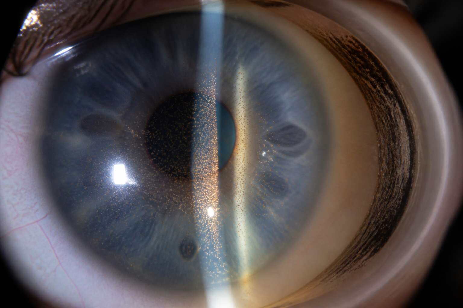
The clinical signs of pigment glaucoma are so characteristic that an experienced examiner can often identify the condition before measuring the pressure. Unlike primary open-angle glaucoma, where the drainage angle looks open yet the resistance remains unexplained, this disorder reveals itself through distinctive physical findings:
- Radial iris transillumination defects where pigment has been rubbed away
- A Krukenberg spindle of pigment dust on the corneal endothelium
- A dense, dark band of pigment within the trabecular meshwork
These findings change the clinical conversation. Pigment glaucoma has a clear anatomical source and a treatment logic that follows from it. Management diverges sharply from standard glaucoma care because the obstruction can sometimes be addressed directly, and because the underlying pigment dispersion syndrome requires its own surveillance. Few glaucoma subtypes offer such a coherent picture!
The Role of Pigment Dispersion in Eye Pressure
Pigment glaucoma begins when the iris bows backward, causing its pigment epithelium to rub against the lens zonules. This friction releases melanin granules into the aqueous humor. These granules accumulate at thedrainageangle, clogging thetrabecularmeshwork. Elevated intraocularpressurefollows, and thehallmarksignsemerge.
Severalstepsdefinethiscascade.
- Theirisbowsbackwardduetoa reversepupillaryblock.
- Pigmentgranulesshedfromtheirispigmentepitheliumontozonulefibers.
- Granulesflowin theaqueoushumortowardthetrabecularmeshwork.
- Meshworkobstructioncausespressurespikes.
Thesestepsarenotrandom;theyemergefromafixedanatomicalrelationship.Patientsoftenaskwhytheiririsshedspigmentsoreadily.Thisdisorderhasadistinctepidemiology,generallymiddle-agedmen,oftenwithmyopia.Thisisafinitemechanicalevent,notaninfectiousprocess.Understandingthereoleofpigmentdispersionineyepressuremeanstrackingthestructurallogicbehindthisgradualshedding,whichsetspigmentglaucomaapartfromotherglaucomas.
Who Is at Risk?
Age and Gender Patterns
For every three men who develop pigment glaucoma, only one woman receives the same diagnosis. This disparity is not a footnote. It is the defining rhythm of the condition. The pathology begins with pigment dispersion, a process that does not discriminate. However, the resulting pressure elevation shows a distinct preference for the male eye.
I continue to find this pattern striking in clinical practice. Men in their late twenties and thirties form the primary cohort. Their deeper anterior chambers allow the iris to bow backward, rubbing against the lens zonules. This mechanical friction releases pigment granules, which then clog the trabecular meshwork. Women, by contrast, typically have shallower anterior chambers, offering a degree of anatomical protection.
Age shapes the narrative as much as gender does. The condition tends to declare itself early, often between the ages of 20 and 40. This is unusual among glaucoma types, which usually emerge later in life. Over time, the pigmentary process can exhaust itself. The iris becomes less flexible, and the eye essentially self-corrects the rub. Yet the damage to the drainage system often persists.
In South Africa, the demographic profile appears consistent with global data, though awareness remains low.
The classic pigment glaucoma patient profile includes:
The age pattern extends into the early forties, where diagnosis peaks before the pressure begins to stabilize.
Genetic and Hereditary Links
Genetics matter more in pigment glaucoma than many patients realize. A first degree relative with pigment dispersion syndrome raises your own risk of developing pigment glaucoma considerably. The condition follows an autosomal dominant pattern in some families, meaning one altered gene from either parent can pass along the vulnerability.
Researchers have identified mutations in the PMEL gene and others that influence iris structure and pigment production. These markers do not guarantee disease, but they increase susceptibility. South African families often discover the link only after one member receives a diagnosis.
Consider these hereditary warning signs:
- A parent or sibling diagnosed with pigment dispersion syndrome
- Multiple family members with unexplained high eye pressure
- Ancestry that includes known glaucoma clusters
Genetic testing remains uncommon in routine eye care, yet a careful family history stays the simplest screening tool in my experience.
Anatomical Features That Increase Susceptibility
The architecture of the eye dictates your vulnerability more than any lifestyle choice. For pigment glaucoma, the critical variable is the anterior chamber’s depth and the peculiar geometry of the iris. When the iris bows backwards, it scrapes against the lens zonules, releasing pigment that clogs the drainage system. This is not a random event. It is a structural flaw, a physical collision happening within a confined space.
Patients with a deep anterior chamber and a concave iris contour carry the highest susceptibility. This specific configuration is often visible during an eye exam, though it rarely causes symptoms.
– A pronounced, concave iris curvature
– An unusually deep central anterior chamber
– A shorter axial length combined with a thicker lens
These features create the friction that accelerates pigment dispersion. Younger men with myopia face the highest anatomical risk, as the eyeball’s elongation stresses the iris-lens relationship. Over time, as the lens thickens with age, the space narrows and the friction often lessens, which explains why pigment glaucoma can stabilize later in life. Understanding your own ocular anatomy is the first step toward recognizing the threat.
Lifestyle and Environmental Influences
I have seen patients dismiss their eye pressure as a random misfortune, yet the pattern is often clear in their daily routines. Strenuous exercise, especially jarring activities like mountain biking on uneven South African terrain, can shake pigment from a vulnerable iris. Similarly, hours of near work, such as embroidery or soldering, alter lens thickness and increase iris friction. Environmental light changes, from deep shade to harsh sun, force rapid pupil responses. Consider these common influences:
- Vigorous cardio with repetitive head movement
- Sustained reading without adequate ambient light
- Abrupt transitions between shade and sunlight
Each encounter is harmless alone, but repeated across months, they push intraocular pressure higher. For pigment glaucoma, the risk accumulates through daily activity, while stillness offers little protection.
Recognizing Symptoms and Getting a Diagnosis
Common Visual Disturbances
Pigment glaucoma rarely announces itself with a single dramatic event. Instead, it tends to show up as intermittent visual noise that you might shrug off. One moment your vision is crisp, the next it blurs after a workout or a long walk. People describe the most common visual disturbances differently, but a few patterns stand out.
- Blurring or fluctuation after physical exertion
- Rainbow halos around bright lights
- Gradual reduction of peripheral vision
These symptoms can come and go, which makes diagnosis tricky. I have seen patients dismiss them as fatigue or stress. An eye care professional can perform gonioscopy to inspect the drainage angle and use visual field testing to map any loss. The condition can progress without obvious pain, so early detection matters.
Physical Signs During an Eye Exam
The diagnosis of pigment glaucoma often begins with what the eye care professional sees before you say a word. A slit lamp examination reveals physical markers that patients cannot feel or notice themselves:
- Krukenberg’s spindles, vertical pigment deposits on the inner cornea
- Transillumination defects, gaps in the iris where pigment has eroded
- A darkened trabecular meshwork visible through gonioscopy
These findings carry diagnostic weight. The pigment dispersion pattern in pigment glaucoma is distinctive, and when paired with elevated intraocular pressure, the diagnosis becomes clear. Gonioscopy allows the clinician to inspect the drainage angle directly, confirming whether pigment has accumulated in the meshwork. The examination is painless and takes minutes, yet it provides the certainty that symptom-based assessments cannot. For many patients, the first concrete evidence of the condition arrives in the examination room, not through any symptom they have experienced.
Diagnostic Tests and Imaging Techniques
Most patients with pigment glaucoma will never feel the damage accruing behind their eyes. The condition progresses without warning, and by the time symptoms force a visit to a clinician, significant visual field loss may already be permanent. Diagnosis therefore depends on a battery of tests that reveal what the patient cannot perceive. Diagnostic tests for pigment glaucoma typically include:
1. Tonometry to measure intraocular pressure.
2. Optical coherence tomography (OCT) to map retinal nerve fibre thickness.
3. Visual field testing to document peripheral vision loss.
These imaging techniques demand precision. A single reading rarely tells the whole story, so clinicians repeat measurements over months, watching for subtle shifts. The diagnosis emerges from patterns, not isolated numbers. For those in South Africa, access to such equipment varies by region, but the underlying principle remains constant: pigment glaucoma hides in plain sight, and only systematic testing pulls it into the open.
The Importance of Early Detection
Treatment and Management Strategies
Prescription Eyedrops and Medications
Treating pigment glaucoma rarely starts with surgery. Prescription eyedrops and medications remain the first line of defense. These drugs lower intraocular pressure, which is the main target. However, pigment glaucoma presents a unique challenge: the pigment granules themselves can obstruct the trabecular meshwork, so pressure control needs consistent monitoring.
I usually recommend a combination approach. Doctors in South Africa often prescribe:
- Prostaglandin analogs like latanoprost to increase fluid outflow
- Beta blockers such as timolol to reduce fluid production
- Carbonic anhydrase inhibitors like dorzolamide for adjunctive pressure control
The choice depends on how much pigment is present and how reactive the eye is. Regular follow-up is essential because medication effectiveness can shift as pigment dispersion continues. Adjusting doses early prevents long-term damage.
Laser Therapy and Its Effectiveness
When eyedrops lose their edge, laser therapy becomes a more decisive step. For pigment glaucoma, the goal isn’t just lowering pressure; it is about preventing the pigment granules from causing further obstruction. I have seen patients where medication simply cannot keep up with the continuous dispersion. In those cases, we turn to the laser.
Selective laser trabeculoplasty, or SLT, targets the drainage channels directly. It does not burn tissue. Instead, it uses a gentle pulse to stimulate the eye’s own healing response. That response clears out the clogged meshwork. We are giving the eye a reset button for its internal plumbing.
– Argon laser trabeculoplasty for more aggressive cases
– Selective laser trabeculoplasty for lighter, targeted treatment
– Laser peripheral iridotomy when pigment blocks fluid flow
The effectiveness varies. Some patients see a significant drop in intraocular pressure within weeks. Others need a second session. The pigment itself can absorb the laser energy differently, so success depends on how the eye reacts. I explain this to my patients clearly, it is not a miracle cure, but for many, it is the difference between stability and a slow slide toward vision loss. Ongoing pressure checks remain non-negotiable after the procedure.
Surgical Options for Advanced Cases
For advanced pigment glaucoma, surgery becomes the primary means of pressure control. Trabeculectomy remains the gold standard in South Africa. The surgeon creates a small passage for fluid to bypass the blocked meshwork, forming a bleb under the conjunctiva. Patients often arrive at this threshold after years of eyedrops and laser sessions. The decision carries weight that goes beyond clinical numbers.
Drainage implants offer an alternative when trabeculectomy fails or the conjunctiva is scarred. The Ahmed and Baerveldt tubes shunt aqueous humor to a reservoir plate. The choice follows a careful assessment of the individual eye.
When surgery is inevitable, the decision depends on:
- Previous surgeries and conjunctival health
- Intraocular pressure targets and disease severity
- Patient age and healing capacity
Success in pigment glaucoma depends on controlling both pressure and continued pigment dispersion. For some patients with pigment glaucoma, combined cataract surgery and trabeculectomy offers a single recovery period.
Long-Term Monitoring and Lifestyle Adjustments
Glaucoma is a chronic condition, and pigment glaucoma demands a particular kind of patience. The disease does not end with a successful surgery or a stabilized eye pressure reading. Instead, the journey shifts into a quieter, more persistent phase of vigilance where the patient’s daily routines intersect with the clinical reality of their diagnosis. For many in South Africa, this means accepting a new relationship with time, where every check-up becomes a small ritual of reassurance.
Long-term monitoring for pigment glaucoma is not a passive act. It requires a commitment to the subtle details that often go unnoticed. The clinical team will track the optic nerve’s appearance, the thickness of the corneal endothelium, and the stability of the visual field over months and years. These measurements form a narrative of their own, revealing whether the trabecular meshwork is coping or if the pigment dispersion continues to exert its relentless pressure. The patient’s own observations, from noticing halos around lights to feeling a dull ache in the eye, become critical data points in this ongoing dialogue.
Lifestyle adjustments weave themselves into this clinical framework. The patient learns to recognize the physical activities that spike intraocular pressure, such as inverted postures or high-impact exercise, and modifies them without losing the joy of movement. Hydration and caffeine intake often come under scrutiny, as both can influence the ebb and flow of aqueous humor. The broader goal is stability, a quiet equilibrium that supports the work done by medication or surgery.
– Maintain a consistent schedule for eye pressure checks, even when symptoms are absent.
– Carry a list of current medications and allergies at all times for emergency visits.
– Protect the eyes from direct trauma during sports or manual labour, as pigment liberation can spike pressure.
– Communicate changes in vision or discomfort to the ophthalmologist without delay, as early intervention alters the outcome.
The psychological weight of this condition is real, but it does not have to define the patient’s existence. With a solid partnership between the patient and their eye care provider, the long-term outlook for pigment glaucoma in South Africa is one of managed stability rather than inevitable decline. The routine becomes a form of empowerment, a daily practice that turns anxiety into action.
Potential Complications and How to Avoid Them
Treating pigment glaucoma is a balancing act between pressure control and preserving the eye’s inner lining. Most patients in South Africa respond well to a stepped approach that begins with medication and advances only when needed. But the disease can complicate quietly. The trabecular meshwork may scar, corneal endothelial cells may drop, and visual field loss can accelerate if pressure spikes go unnoticed.
Avoiding these outcomes requires honesty during follow-ups. Patients who report exercise habits, caffeine intake, or missed drops give the ophthalmologist a true picture. Without that candour, adjustments happen too late.
- Track pressure checks consistently
- Report halos or pain immediately
- Wear protective eyewear during sport
No treatment erases the pigment glaucoma. It manages it. That distinction matters for every long-term plan.
Living With the Condition
Daily Tips for Protecting Your Vision
Emotional and Psychological Support
The creeping of pigment glaucoma can unsettle a person’s certainty. This condition often arrives amidst routine appointments, and the follow ups may stir unexpected loneliness. Many individuals attend many tests and pressure checks while their inner state holds persistent worry.
Emotional support for those living with pigment glaucoma comes from several directions. A shared conversation with a trusted friend, a support group, or a psychologist can ease the strain. In South Africa, urban centres like Johannesburg and Cape Town host glaucoma societies that offer fellowship. These spaces allow people to voice fears without shame, and the recognition of shared experience brings comfort.
Common feelings that emerge include:
- Frustration at repeated clinic visits
- Fear of losing independence
- Relief when a pressure reading stabilises
Understanding these emotions matters as much as any medication. The psychological impact continues through daily life, yet acknowledging it strengthens resolve.
Resources for Patients and Caregivers
Living with pigment glaucoma demands a patience that no one volunteers for. In South Africa, where specialist care can be hours away, the distance between diagnosis and daily life feels significant. For patients and caregivers, practical resources matter as much as clinical ones. Patient education booklets from ophthalmology departments help translate pressure readings into daily understanding. Caregivers, who hold their own concerns, can find guidance through hospital social workers and community health clinics.
- The Glaucoma Society of South Africa offers patient days and printed guides
- Regional hospitals in Gauteng and the Western Cape run low vision clinics
- Online forums connect rural caregivers with urban specialists
These tools do not erase pigment glaucoma, but they make the path clearer for patients and caregivers alike. That clarity, I have learned, matters more than most people expect.



0 Comments